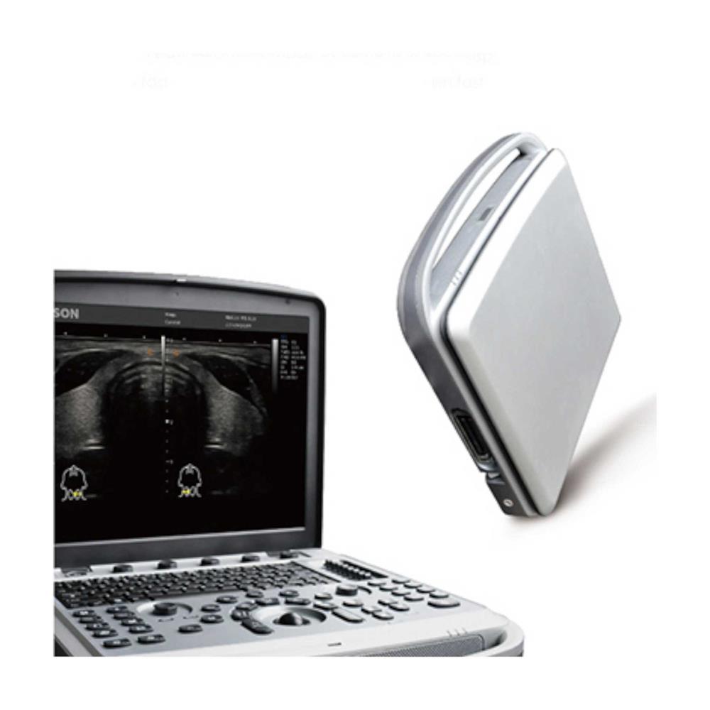
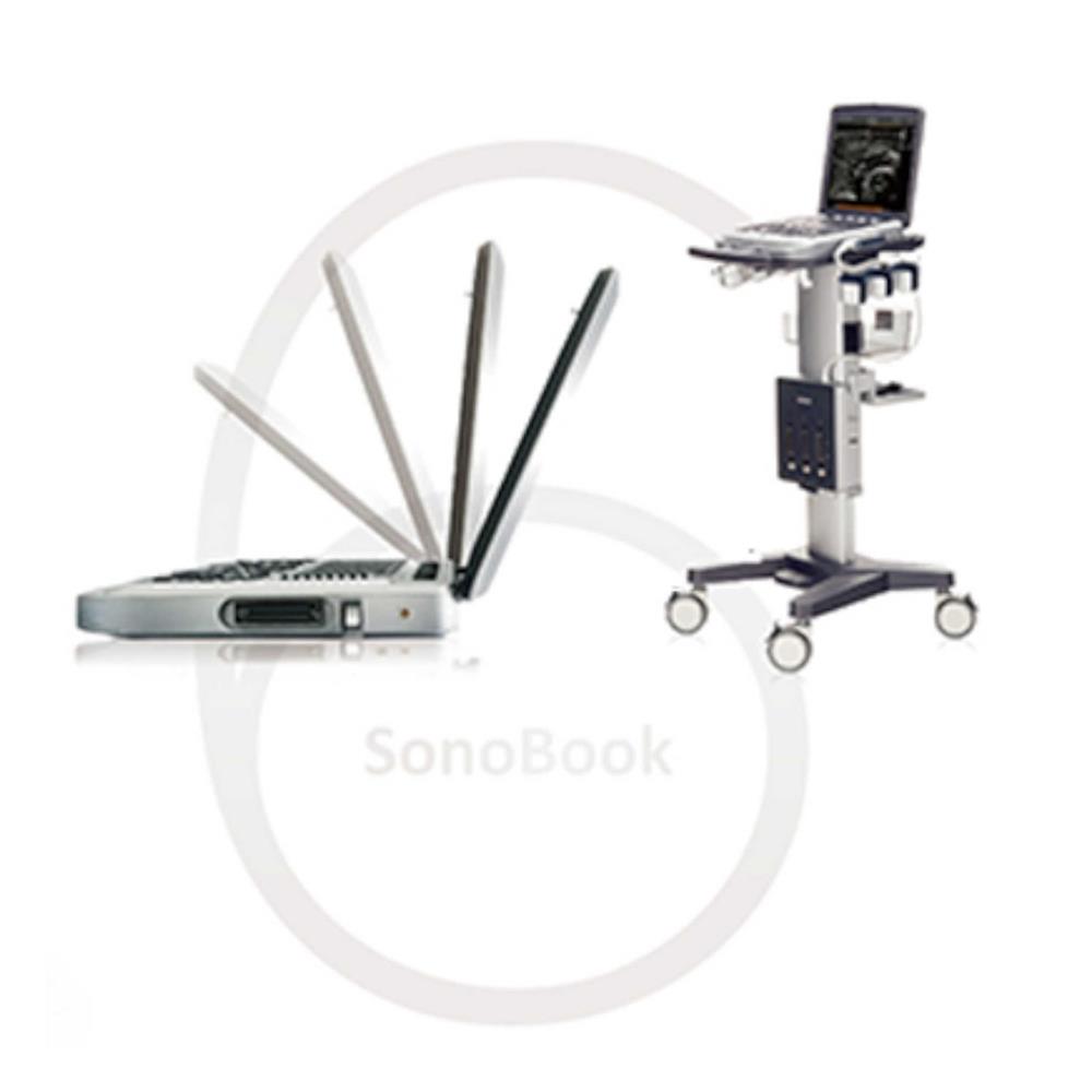
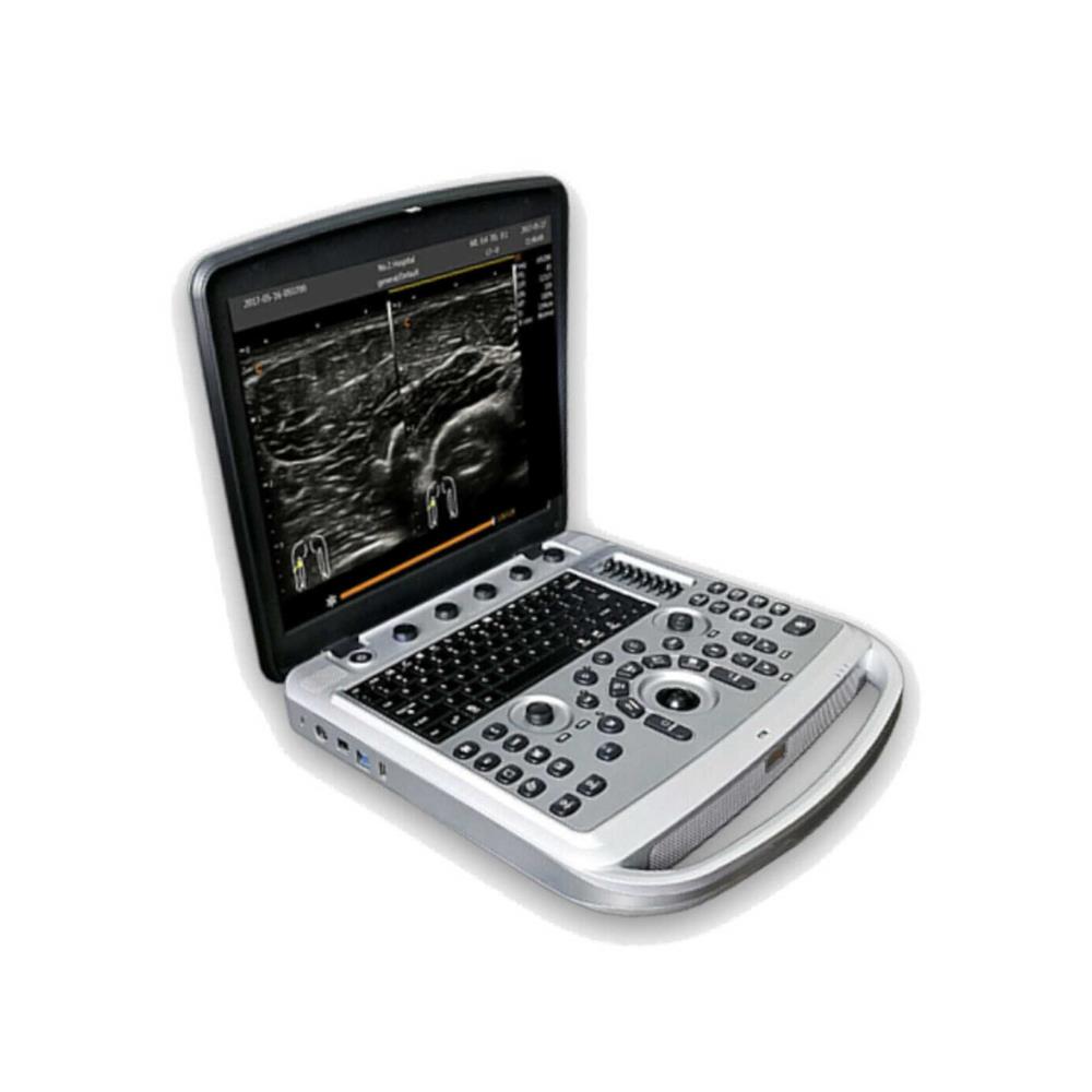
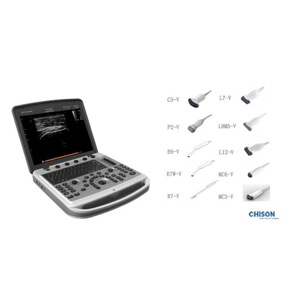
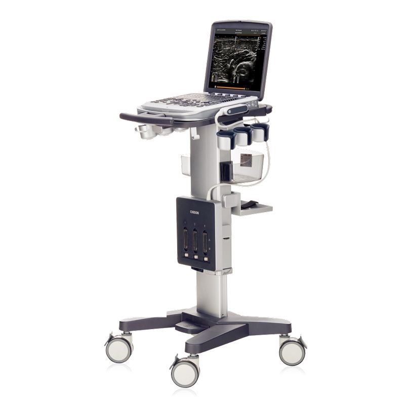
Color ultrasound SonoBook 6 Main unit

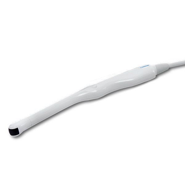
***Does not include the probes.

- Description
Description
Chison SonoBook 6 ultrasound
The new Chison SonoBook 6 portable, features with compact laptop-based design for easy mobility, to provide solutions when fast reaction is needed.
Combined with productive features, crisp imaging performance and intuitive workflow while offering economic clinical value.
It has advanced features such as: Compound Imaging, Speckle Reduction Imaging, SuperNeedle (Needle Visualization), Elastography, AutoIMT, Tissue Doppler, HIP Graph, and full DICOM with Structured Reporting.
Its internal battery allows for up to 2 hours of scan time.
Features:
Imaging Modes 2D, Auto Optimization, Color Doppler, AutoIMT, Compound Imaging, DICOM, IMT, M-Mode, Power Doppler, PW Doppler, Speckle Reduction, Tissue Harmonics.
Applications:
Emergency Medicine, Anesthesia, Breast, Small Parts, Superficial, Musculoskeletal, OB/GYN, Abdominal, General Imaging, Vascular, Pediatrics.
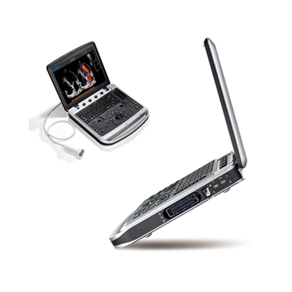
15" LED HD monitor, 120° rotatable
Truly portable design, less than 6 kg.
Longer battery life up to 2 hours active mode, fast battery swap.
Triple probe connector and precision cart(optional).
SonoDocking: DVI-I, Foot-switch, Video-out, remote, S-Video, for fast data transfer.
Video and Output Options: USB, Ethernet, DVI, S-Video.
Dimensions: 12 lbs, 14? x 14? x 3?.
Export Options: USB, BMP, JPG, AVI, Network Storage, DICOM.
USB Ports: 2.
DICOM Options: Store, Print, Worklist, MPPS, Structured Reports.
PC Export Formats: BMP, JPG, AVI.
CD/DVD-R: N/A.
Ethernet: Yes.
Wireless: Yes.
Power (USA): 100-240V, 50/60 Hz.
Battery: Yes.
HDD Size: 320GB.
SSD/HDD: HDD.
Max Cine Memory: 256 Frames.
Maximum Depth: 30 cm.
Productive features:
Elastography:
Display the elasticity of different tissues in different color.
Provide more clinical information especially for beast tumor, thyroid, liver and prostate.
Strain ratio measurement, quantitatively gives the ratio between the average strain of the selected region and the nearby normal tissue region.
Auto IMT:
Super Needle:
Enhance the needle displaying in image, without distortion of the needle.
Decrease the complicated rate, increase the successful rate.
HIP Graf:
Use the graph for hip orthotics diagnosis, help the doctor to give an easier and more accurate diagnosis during the pediatric hip scanning.
Different angle indicate different level of hip deformity, which is easy and obvious to see with the aid of the graph. (I, II, D, IIIa, IIIb)
Intuitive workflow:
Six-one-key Procedure.
Anatomy-specific presets.
Efficient Data Management.
User-friendly interface.
Shortcut key for quick access.
Auto measurements.
Complete DICOM function.