Deluxe demonstration skull model 14-part for advanced studies
Top quality skull model
- Description
Description
Deluxe demonstration skull model 14-part for advanced studies
Excellent quality model that faithfully copies a real human skull and depicts all anatomical structures in the highest detail.
It is ideally designed for the education of students in the fields of anatomy, medicine, surgery, otolaryngology, ophthalmology, and dentistry.
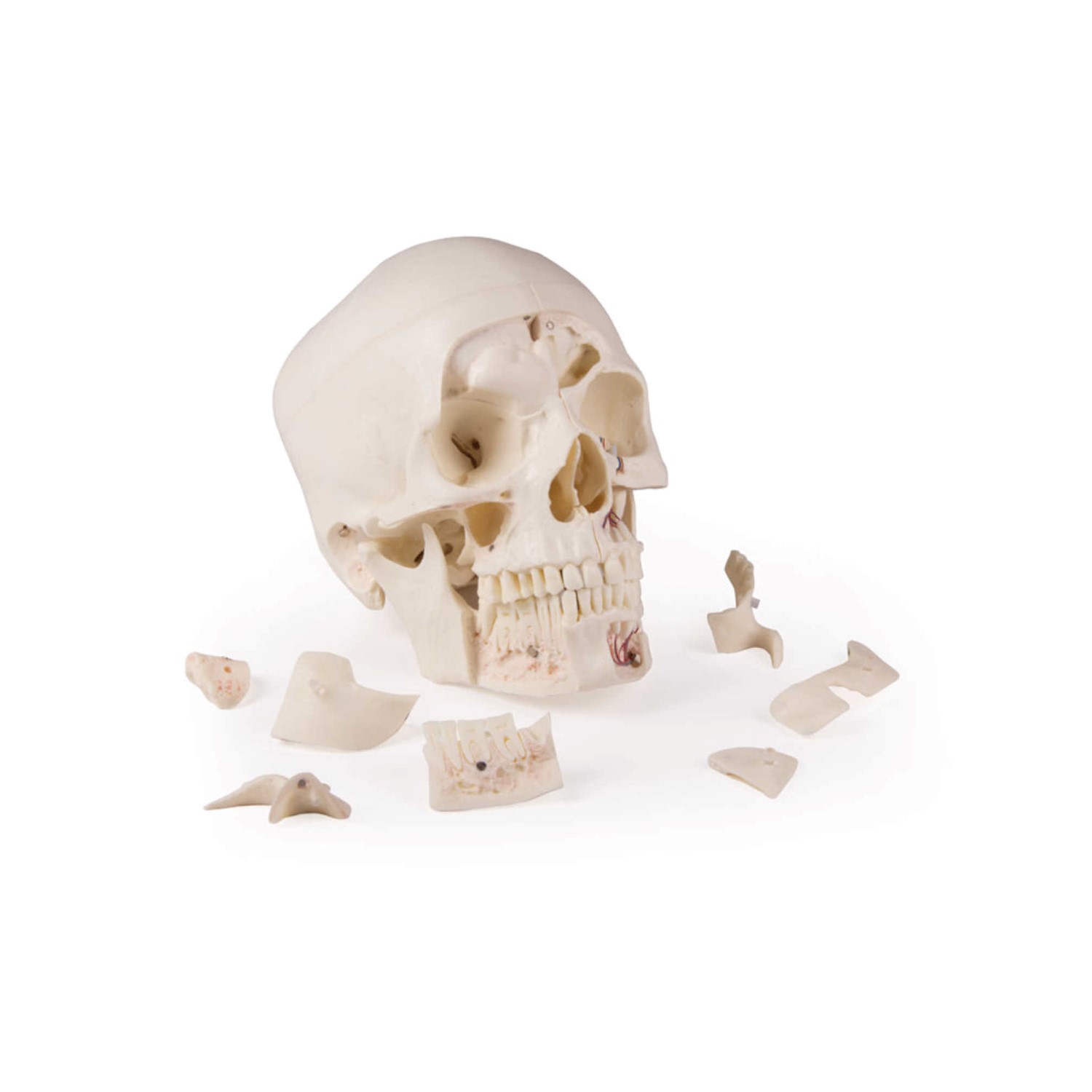
The skull has metal and magnetic connections so that it can be disassembled and fully recovered with ease and comfort.
The model has a horizontal incision, leaving the temporal bone and its connections intact.
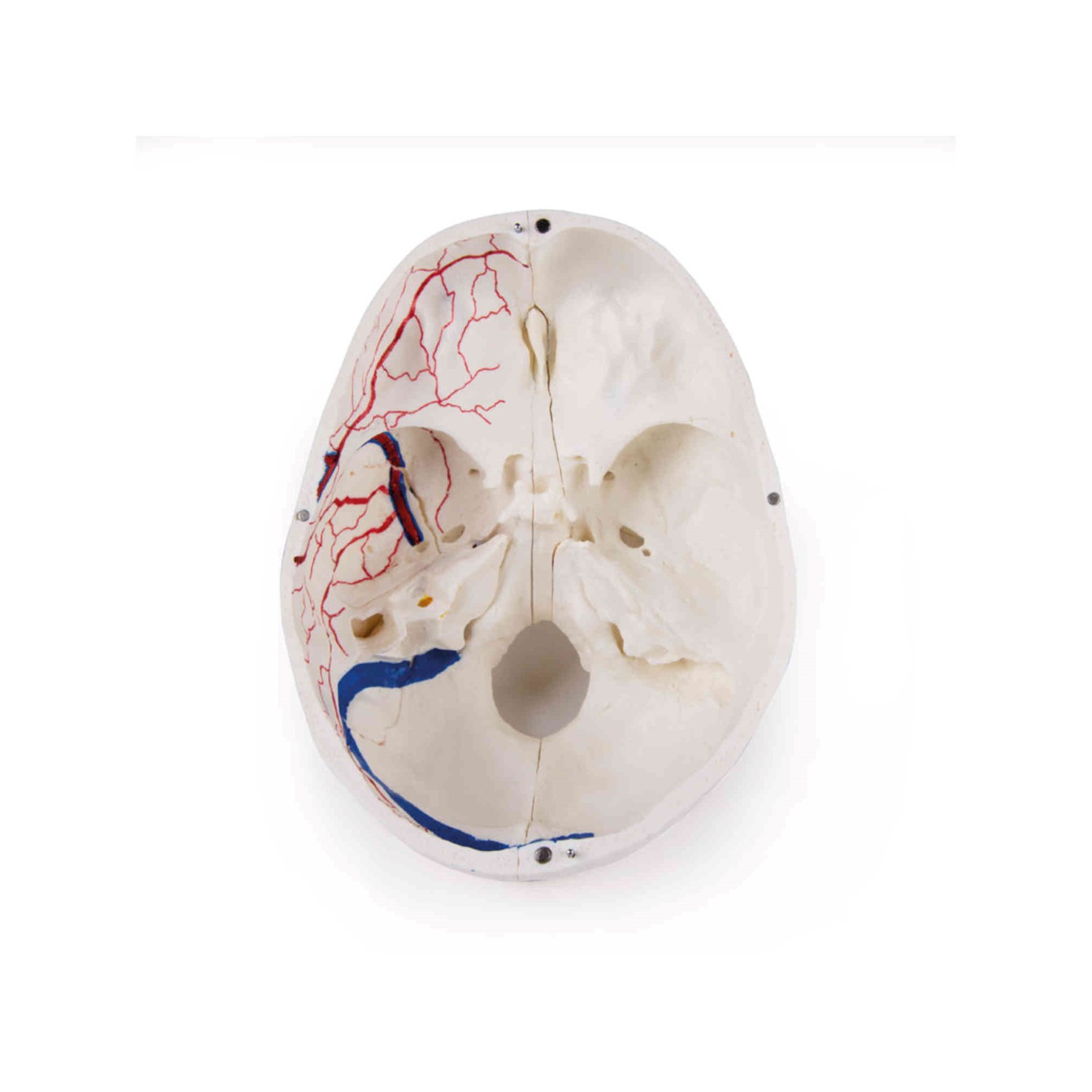
In addition, the bone scans of the upper sagittal sinus, the transverse sinus and the sigmoid sinus, as well as the meningeal vessels, have been painted.
The main part of the skull is split apart, leaving the nasal septum intact as well as the entire nasal septum.
The structures of the anterior, middle and posterior cranial holes are easily accessible.
The nasal cavity, the orbit, the nasal septum, the pharynx and the nasopharyngeal space are also shown.
The nasal septum is separable from the surrounding bones.
The frontal sinuses have been analyzed on one side to show the sinus as a whole and on the other side are carved for full access to the area.
The relationship of the frontal area with the nasal cavity is clear, so it is especially valuable for otolaryngologists.
On one side of the skull the temporal bone has been left in place.
The other temporal bone is removed from the skull.
The three (3) semicircular canals are visible along with the course of the facial nerve that turns backwards and then downwards, where it eventually emerges through the stylo-mastoid foramen.
The removable temporal bone has the external auditory canal intact.
The carotid canal can be opened like the cochlea, showing the internal canal and there is an illustration of the course of the facial nerve.
The semicircular canals and the area of ??the tympanic orifice are visible.
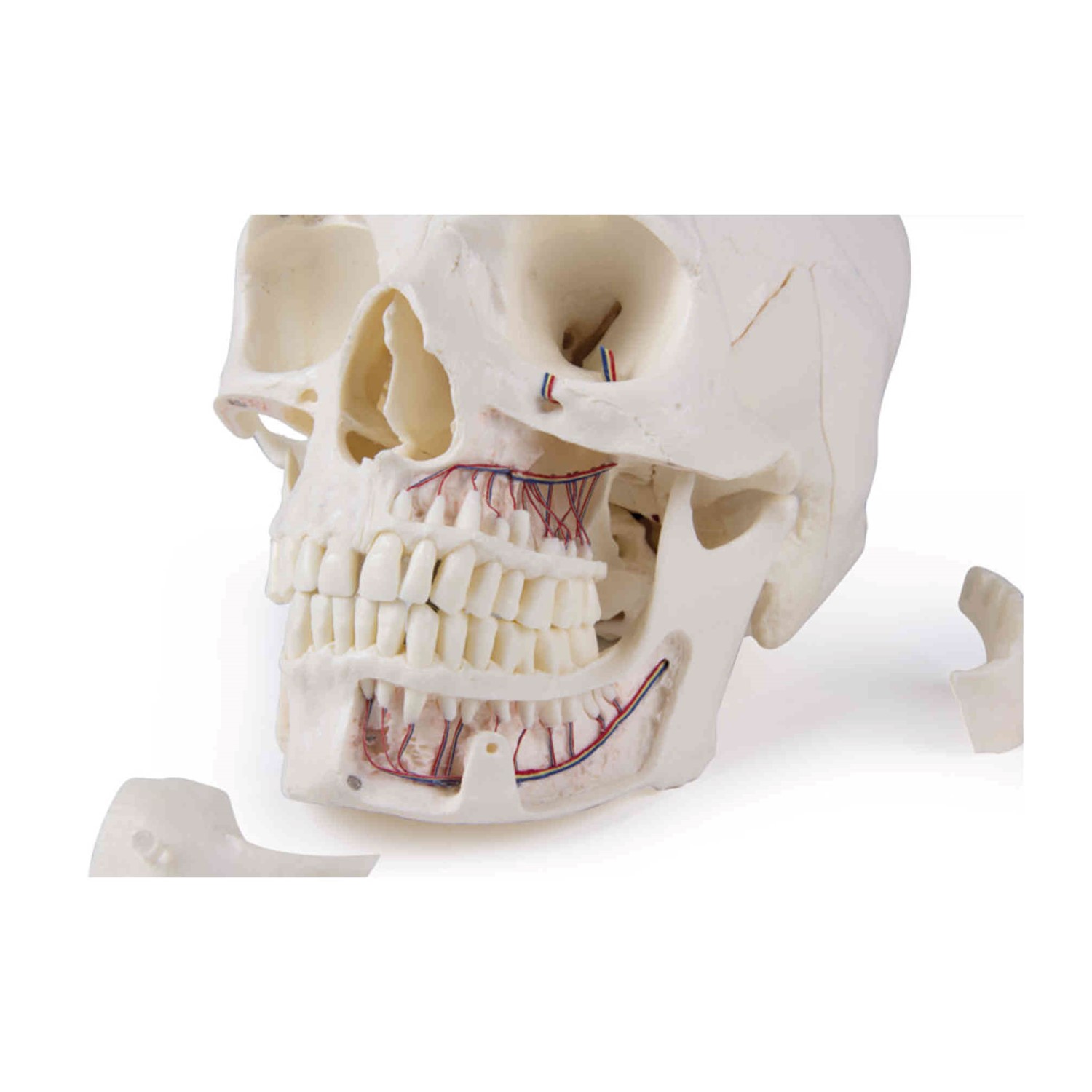
The upper jaw and lower jaw expose the structures of the dentition, the roots, the dental vessels and the nerves are visible.
The maxillary region can be opened by removing part of the bone.
The teeth of the right jaw have incisions to reflect the internal structure of the tooth.